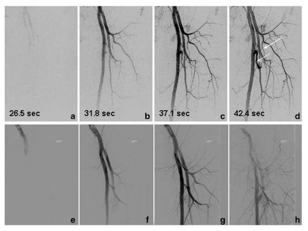
Figure 3.
Results from a time-resolved study of Patient 11, a 46-year-old male with a vascular malformation of the left upper leg. Consecutive targeted MIPs (a–d) show feeding vessels to the nidus (short arrow). The time series portrays feeding of the lesion by means of the circumflex femoral artery (long arrow). Every other image from the from fluoroscopic angiography series (e–h) performed at the time of treatment that show excellent correlation with the CAPR time series.

