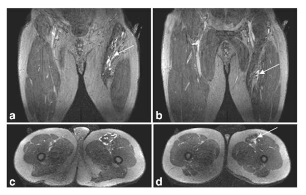
Figure 4.
Coronal (a,b) and axial (c,d) slices from the late phase imaging in Patient 11 shows that the lesion is restricted to the sartorius muscle, with the muscle compartment outlined (c, dashed yellow box). (a) and (c) depict the more proximal portion of the lesion and (b) and (d) the more distal part of the lesion.

