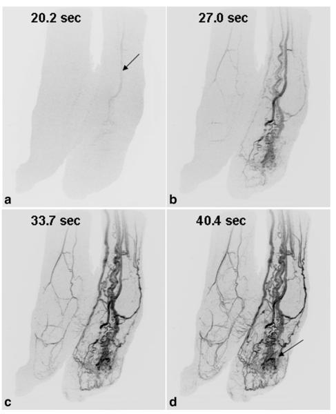
Figure 5.
Results from Patient 7, a 56-year-old woman with a vascular malformation of the left foot (a–d). Oblique MIPs of both feet show rapid contrast passage through the enlarged vessels of the left foot compared with the right foot. Dominant supply to the malformation is from the anterior tibial artery (arrow, A). The time series also shows involvement of the malformation with the plantar skin of the foot (D, arrow).

