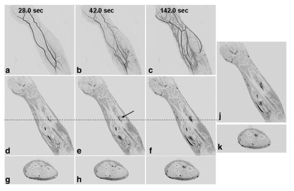
Figure 6.
Results from the CAPR time-resolved series and late phase imaging from Patient 12, a 34-year-old male with a vascular malformation of the left forearm. Coronal MIPs (a–c), coronal slices (d–f), and axial slices (g–i) from the time-resolved series with a 3.5-s frame time show progressive arterial and venous filling with late enhancement of the malformation. Due to the enhancement of the superficial veins, it is difficult to identify the enhancement of the malformation in the MIP images. The malformation is more clearly seen in the slices. Coronal and axial slices from the last time-resolved image (f,i) show similar depiction of the malformation as the late single phase image (j,k).

