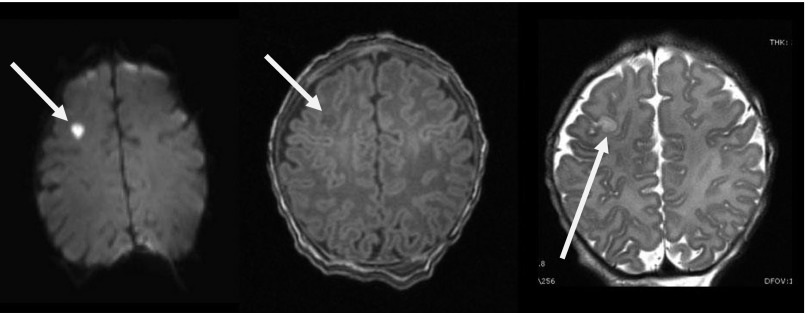
FIGURE 1.
Stroke on MRI scan. A right cortical stroke (white arrow) identified by a high-signal intensity on DWI (left), a low-signal intensity on T1-weighted imaging (middle), and increased signal intensity on T2-weighted imaging (right) on the MRI scan of an asymptomatic newborn. This finding on MRI resulted in delay of surgery for this patient who had a normal HUS.

