
Figure 4.
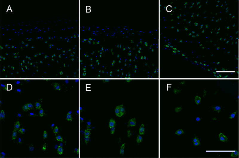
Effects of cytochalasinB and nocodazole on chondrocyte cytoskeleton. Confocal images showing phalloidin stained f-actin (green) and DAPI stained (blue) bovine cartilage of cytochalasinB (A,D), nocodazole (B,E), and untreated (C,F) specimens. CytochalasinB and nocodazole groups show dissociated f-actin in the superficial zone whereas the untreated group has intact f-actin. Scale bars in C and F are 100 and 50 microns, respectively.