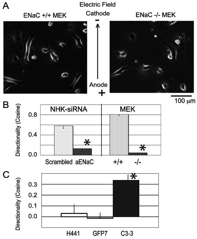
Fig. 1.

Depletion of ENaC inhibits galvanotaxis of keratinocytes, but overexpression of EGFP-ENaC promotes galvanotaxis. (A) Representative images from the wild-type MEK and the ENaC-depleted MEK in EF (also supplementary material Movies 1–4). (B) The migration speeds of keratinocytes are between 1 and 2 µm/minute. Left: The directionality decreases by 80% in human NHK treated with αENaC siRNA (120 cells tracked). Right: αENaC-KO-MEK also lose 94% of their directionality in the EF (+/+ wild type, 104 cells tracked; −/− αENaC knockout, 93 cells tracked). Although αENaC-KO-MEK migrate faster than wild-type MEK, the migration speed of the ENaC siRNA-treated NHK is similar to that of scrambled siRNA-treated NHK (1.2±0.46 µm/minute versus 1.1±0.47 µm/minute; not significantly different; migration speed not shown). (C) Galvanotaxis of parental H441 cell lines H441 (80 cells tracked), GFP7 (EGFP expressing H441, 165 cells tracked) and αC3-3-GFP (EGFP-αENaC expressing H441, 165 cells tracked) were examined. The H441 and GFP7 lines that express very low levels of endogenous αENaC do not respond with directional migration to the EF (cosine is close to zero). However, introduction of a functional αENaC channel (in the αC3-3-GFP) significantly increases the cathodal directionality (cosine) of the cells. *P<0.05.
