Fig. 4.

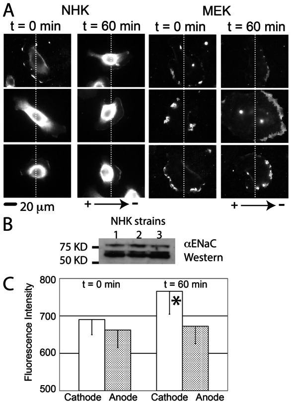
αENaC polarizes to the cathodal side of keratinocytes after 60 minutes of galvanotaxis. (A) Immunostaining shows that αENaC is distributed randomly at cell periphery of NHK and MEK before exposure to the EF (0 minutes, cathode on right of the image). After 60 minutes in the EF, ENaC is concentrated at the cathodal side of cells. (B) Both uncleaved (∼80–85 kDa) and cleaved αENaC (60–65 kDa) bands were detected with western blotting on three strains of NHKs. (C) At 0 minutes, the fluorescence intensity between the cathodal side and anodal side of keratinocytes is similar (left), but at 60 minutes, the fluorescence is increased at the cathodal side (right, *P<0.05, 20 human keratinocytes analyzed in each group).
