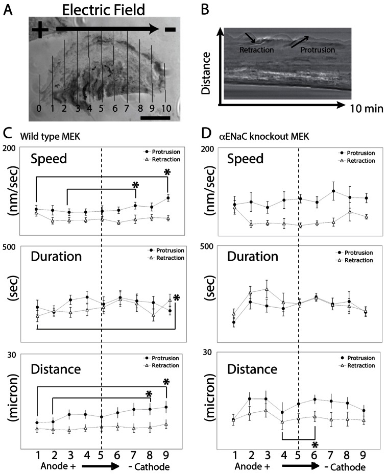
Fig. 5.

ENaC is required to establish stable lamellipodia at the cathodal side of galvanotactic keratinocytes. (A) Mouse keratinocytes were exposed to the EF and filmed for 10 minutes. Fan-shaped cells were selected and a 1-pixel wide line at cell periphery was drawn every 10% of the length (lines were aligned from anode to cathode, numbered 0–10) across the MEK to generated kymographs. (B) The protrusion and retraction of the lamellipodia were tracked and plotted from either wild-type MEK or αENaC-KO-MEK. (C,D) Wild-type MEK lamellipodia protrude faster and further at the distal cathodal side compared to the anodal side (C, n = 13). The asymmetric protrusion is ENaC-dependent and the lamellipodia extended at a similar rate and to a similar distance at the distal sides in the αENaC-KO-MEK (D, n = 7). Quantitative data are presented in Table 1. *P<0.05 for the comparisons indicated.
