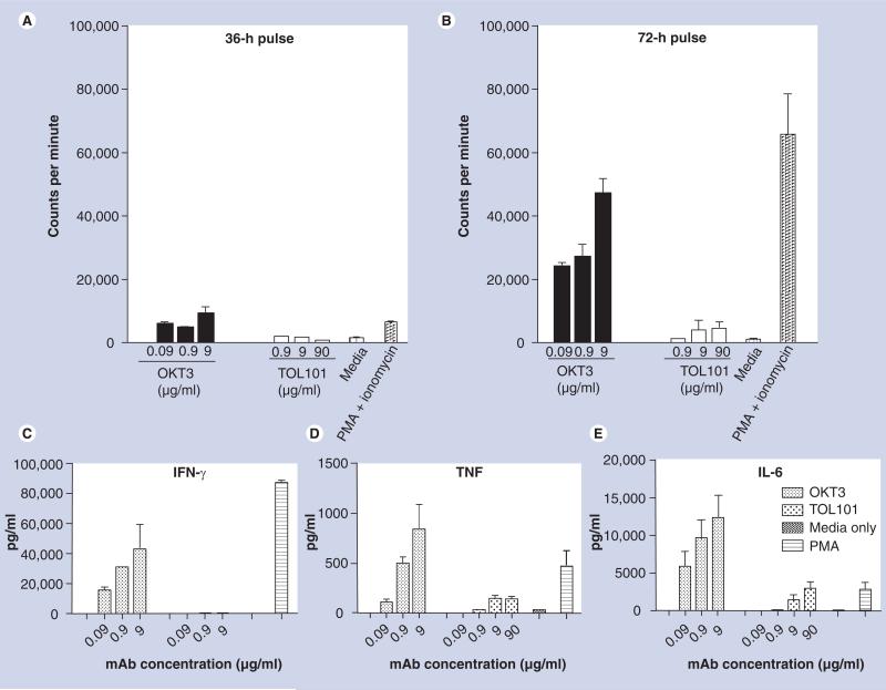
Figure 2. In vitro proliferation assays using freshly isolated human peripheral blood monocyte incubated with OKT3, TOL101, media alone (negative control) or phorbol 12-myristate 13-acetate and ionomycin (positive control).
After (A) 36 h or (B) 72 h, tritiated thymidine was added to the cultures for 12 h, after which the plates were harvested and the incorporation of thymidine measured as a reflection of the mitogenic capacity of each treatment. While OKT3 and PMA and ionomysin induced significant T-cell proliferation, TOL101 did not. The inability to induce T-cell proliferation was also reflected in a lack of proinflammatory cytokine: (C) IFN-γ, (D) TNF and (E) IL-6. Data presented are the mean and standard deviations from three individual patients’ PBMCs. These experiments have been repeated over three times with over nine independent donors.
mAb: Monoclonal antibody; PBMC: Peripheral blood monocyte; PMA: Phorbol 12-myristate 13-acetate.

