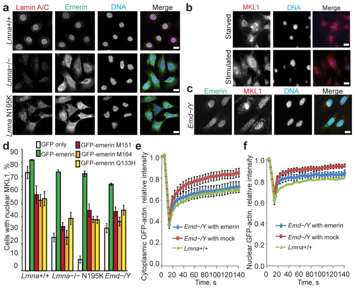
Figure 4. Emerin expression rescues actin dynamics and restores MKL1 nuclear translocation in Lmna−/− and Lmna N195K cells.
(a) Representative immunofluorescence images showing mislocalization of emerin from the nuclear envelope in Lmna−/− and Lmna N195K MEFs. Scale bar, 10 μm. (b) Emd−/Y MEFs had the same defects in MKL1 translocation as lamin mutant cells (compare with Fig. 1a). Scale bar, 10 μm. (c) Stable expression of HA-emerin in Emd−/Y MEFs restored normal nuclear MKL1 localization (71.1±6.02%) in response to serum stimulation. Scale bar, 10 μm. (d) Quantification of nuclear MKL1 localization upon serum stimulation in Lmna+/+, Lmna−/−, Lmna N195K, and Emd−/Y MEFs transiently expressing GFP-emerin, emerin mutants that do not bind to actin (GFP-M151, GFP-M164, GFP-Q133H), or GFP vector alone. Cells were categorized as either having ‘nuclear’ or ‘diffuse/cytoplasmic’ localization of MKL1. Expression of GFP-emerin restored serum-induced nuclear localization of MKL1 in Lmna−/−, Lmna N195K and Emd−/Y cells. N = 50 for each cell line. Statistical significance determined by One-way ANOVA (P ≤ 0.001) with Dunnett Multiple Comparison Post Test. Each group was compared to Lmna+/+ expressing GFP-emerin. (e–f) FRAP analysis of GFP-actin mobility in the cytoplasm (e) and in the nucleus (f) of Emd−/Y MEFs stably expressing either HA-emerin or a mock control. N = 10 for each cell line. Lmna+/+ data reproduced from Fig. 3a for comparison. Error bars, s.e.m.

