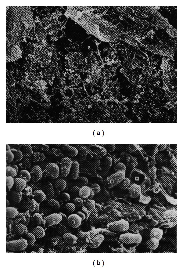
Figure 1.

Propionibacteria in a comedone from acneic skin: interaction with sebum and corneocytes. Scanning electron micrographs of propionibacteria growing on the surface of corneocytes with an open comedone. The image on the right is a higher magnification (×15,840) and shows sebum droplets adhering to the surface of the bacterial cells. Where is the aqueous component? B: bacteria; S: sebum; H: hole in corneocyte. Arrows indicate dividing cells with visible septa. Images reproduced with permission from WH Wilborn, BM Hyde, Montes LF, Scanning Electron Microscopy of Normal and Abnormal Human Skin, 1985, VCH Publishers.
