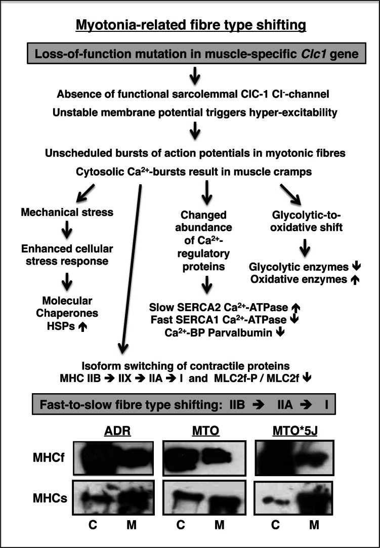
Figure 4.
Effect of myotonia on the expression of skeletal muscle proteins. The flow diagram lists the various steps involved in the molecular pathogenesis of myotonia as revealed by biochemical and proteomic analyses. The immunoblot illustrates expression changes in the fast MHCf and slow MHCs myosin heavy chains in control muscle (C) versus myotonic muscle (M) from mouse mutants ADR, MTO and MTO*5J (36). Immuno-decoration was carried out by standard procedures as previously described by our laboratory (46). Although antibodies to MHCf and MHCs cannot differentiate between the many subspecies of myosin heavy chains, the immunoblot analysis illustrates the general trend of a fast-to-slow switch in isoforms of the contractile apparatus.

