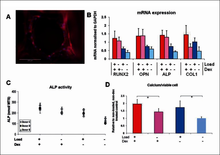
Figure 2.
Compression loading of human MSCs in polyurethane foam scaffolds. A: Fluorescent micrograph of a pore of the scaffold with MSCs attached (blue = cell nucleus stained with DAPI, red = cell cytoskeleton stained with TRITC-phaloidin). B: PCR analysis of mRNA for RUNX2, OPN and ALP showed that these genes were only slightly upregulated by the short (2 hour) loading period and not as much as by continuous dex treatment. However Col 1 was upregulated by loading and inhibited by dex which was also reflected in collagen analysis by Sirius red at a later time-point (data not shown). ALP activity was stimulated by loading to levels seen in dex treated cells as was calcium secretion which was highest with a combination of dex and loading. Adapted from (20) reproduced with kind permission from eCM journal (www.ecmjournal.org).

