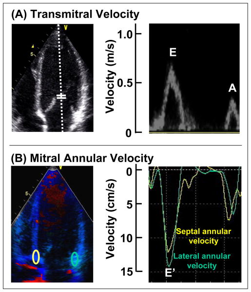
Figure 1.
Examples of echocardiographic data obtained. (A) Transmitral velocities with pulse-wave Doppler in an apical 4-chamber view at the mitral leaflet tips for E and A wave velocity and E wave deceleration time. (B) Mitral annular velocities of septal and lateral mitral annulus (E′) using color tissue Doppler images. Septal and lateral annular velocities were averaged, with E/E′ used as an estimate of left ventricular filling pressure.

