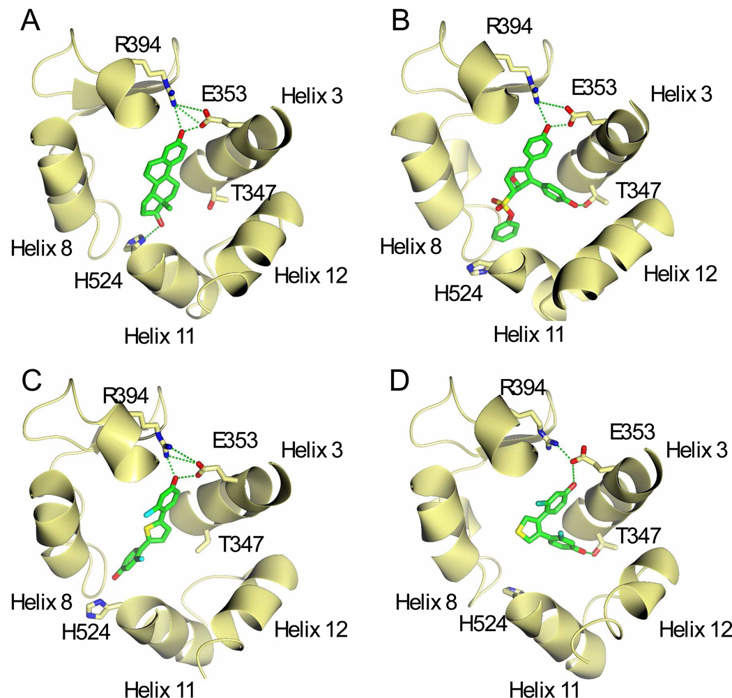
Figure 7. Model of thiophene ligands bound to ERα and comparisons with estradiol and OBHS.
A portion of the ligand binding pocket is rendered as a ribbon, with selected amino acids shown as sticks. (A) Structure of E2 bound ERα. The A ring of E2 forms hydrogen bonds with E353 and R394 while the D ring bonds to H524 (PDB ID: 1ERE). (B) Structure of Oxabicyclic heptane sulfonate (OBHS) bound ERα.52 OBHS H-bonds to both the conserved R394/E353 pair, and also T347 on helix 3. The phenyl sulfonate binds extends between helices 8 and 11. (C) Model of 2e bound to ERα with the conserved H-bonding to R394/E353, and the other phenol extending between helices 8 and 11. (D) Model of 4e with H-bonds to E353 and T347.

