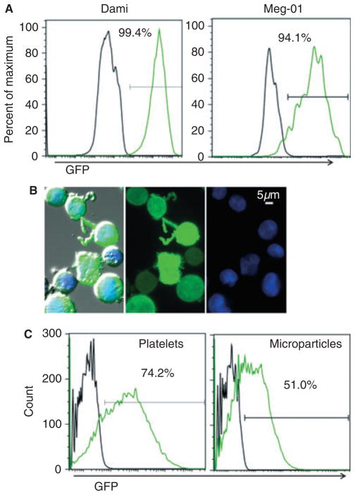
Fig. 1.
Green fluorescent protein (GFP) lentiviral transduction of megakaryocytes results in GFP-positive platelets and microparticles. (A) Two megakaryoblastic cell lines were transduced with a GFP-expressing lentiviral vector. Unstained cells were analyzed by flow cytometry 1 week post-transduction, and GFP fluorescence is shown for untransduced (black line) and GFP-transduced (green line) megakaryocytes. (B) Cytospun GFP-transduced Meg-01cells were stained with the nuclear dye 4′,6diamidino-2-phenylindole (DAPI) (blue). From left to right: the triple-merged DIC bright-field image, GFP fluorescence, and DAPI fluorescence; × 40 objective. (C) Culture-derived platelet-like particles and microparticles were pre-gated by size, and analyzed by flow cytometry for GFP fluorescence. Platelet-like particles and microparticles derived from untransduced or GFP-transduced megakaryocyte cultures are depicted with black and green lines, respectively.

