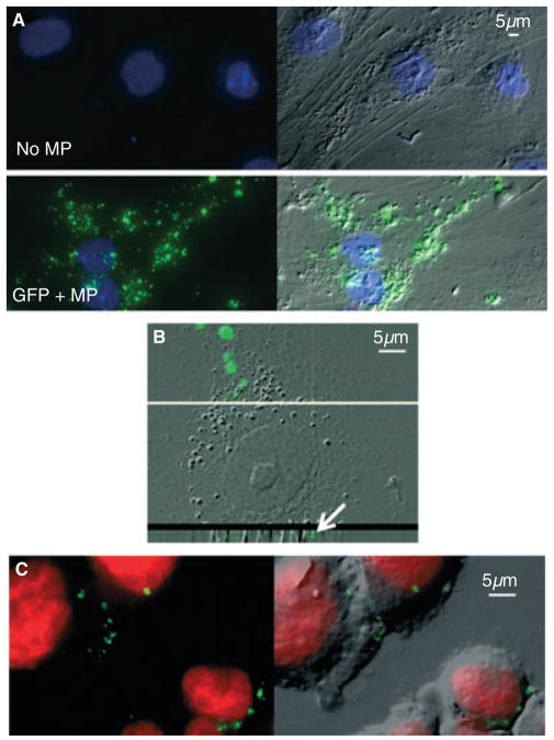
Fig. 3.
Green fluorescent protein (GFP)-positive microparticles are taken up by target cells. (A) Culture-derived microparticles were incubated with preseeded adherent human microvascular endothelial cells (HMVECs) for 6 h. Chamber slides were thoroughly washed before fixation, permeabilization, and 4′,6-diamidino-2-phenylindole (DAPI) staining (blue). Representative examples of HMVECs that were not incubated with microparticles (top) and GFP-positive microparticles (bottom) are displayed as merged GFP (green) and DAPI images (left), and again with the fluorescence overlaying the DIC bright-field image (right); × 40 objective, light microscope. (B) Representative image of confocal microscopy of a GFP-positive microparticle-treated HMVEC. A magnified DIC and GFP merged image of the stacked layers of the cell is shown (top). The horizontal white line shows where the transverse z-stack image is shown below. The white arrow depicts GFP fluorescence within the transverse image of the cytoplasm; × 60 objective, confocal microscope. (C) THP-1 suspension cells were used as other target cells to internalize microparticles. After the cells had been incubated with GFP-positive microparticles for 6 h, they were thoroughly washed before fixation and cytospin centrifugation, followed by Draq5 (red) nuclear staining. Representative images showing GFP and nuclear stain fluorescence (left) are overlaid onto the DIC bright-field image (right); × 40 objective, light microscope. MP, microparticle.

