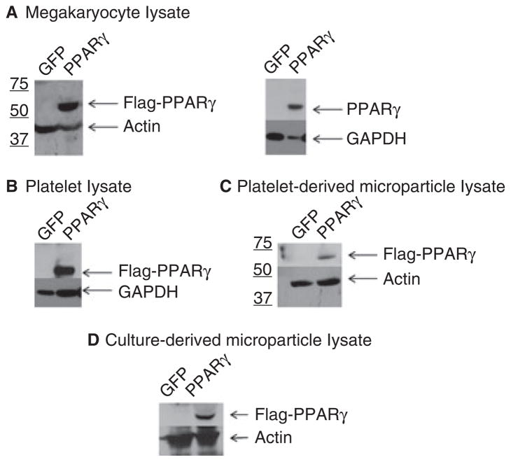
Fig. 4.
Western blot analysis of peroxisome proliferator-activated receptor-γ (PPARγ)-transduced megakaryocyte culture components. Meg-01 megakaryocytes were transduced with either a control lentivirus (green fluorescent protein [GFP]) or a GFP/PPARγ-overexpressing lentivirus (PPARγ). Fractionation of culture components was performed as described in Table 1. Lysates of megakaryocytes (A), platelet-like particles (B), platelet-derived microparticles (C) and culture-derived microparticles (D) were analyzed for protein expression of Flag–PPARγ. Representative western blots were probed with anti-Flag (Flag–PPARγ) or anti-PPARγ (PPARγ) antibodies, as well as actin or glyceraldehyde-3-phosphate dehydrogenase (GAPDH) antibody as the loading control.

