Figure 5.
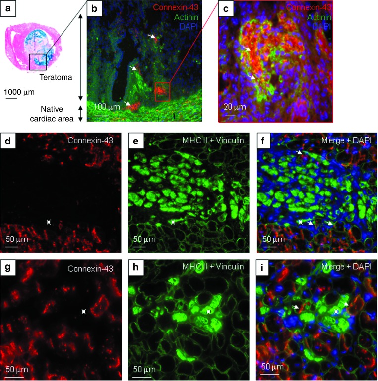
Cardiac differentiation profile of committed ESC and Mb upon transplantation. In the (a–c) Lmna-ESC group, engrafted ESC persisted in two mice as (a) teratomas (stained in blue with X-Gal reagent). Cx43 (red), α-actinin (green), and DAPI (blue) expressions were observed (arrows) in the native cardiac tissue, and in clusters within the tumoral mass. In the (d–i) Lmna-D7 group, Cx43 labeling (red) was observed in the native cardiac tissue, and at the periphery of the newly developed skeletal myogenic tissue stained for MHC II (inside of fibers in green). (e,f,h,i, green) Cardiac and skeletal muscle cells were delineated by vinculin staining. (f,i) Skeletal myogenic myotubes or fibers (green, *) did not express cardiac antigens. DAPI, 4',6-diamidino-2-phenylindole, dihydrochloride; ESC, embryonic stem cells; Mb, myoblasts; MHC, myosin heavy chains.

