Figure 4.
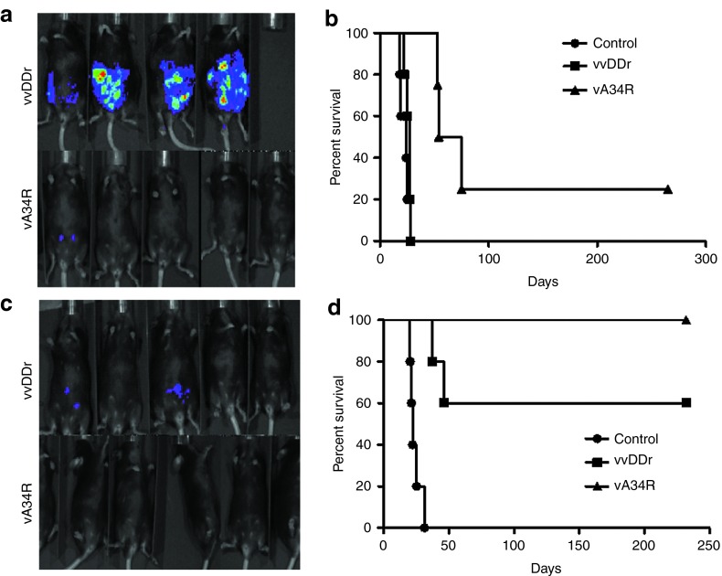
Remote spread in vivo. (a) In vivo bioluminescence imaging for the formation of MC38-luc tumor by uninfected cells when the same were infected with either vA34r or vvDDr at an MOI of 0.001 ex vivo. Tumor burden visualized by luciferase activity in naïve B6 mice, 7 days after i.p. injection of 1.0 × 10⁶ MC38-luc cells infected ex vivo with either virus at an MOI of 0.001 or mock infected (control). (b) Kaplan–Meier analysis of these mice revealed that vA34R cohort developed PC slower and less often, thus resulting in prolonged survival compared with the vvDDr cohort, all of which developed PC (median survival, 54 versus 25 days, P = 0.002) and mock-infected mice (median survival, 54 versus 19 days, P = 0.00184). There was no statistical difference between the vvDDr and control cohorts. (c) In vivo bioluminescence imaging in preimmunized mice in both vvDDr and vA34R groups where the viruses are infected ex vivo in MC38-luc cells at an MOI of 0.01. (d) Kaplan–Meier analysis of these VV-immunized mice after i.p. injection of 1.0 × 10⁶ MC38-luc cells infected ex vivo with vvDDr or vA34R at an MOI of 0.01 or mock infected (control). Median survival of vvDDr group and vA34R group (>232 days) were longer than the mock-infected group (25 days) in a statistically significant fashion (log rank test, P = 0.003). MOI, multiplicity of infection.

