Fig. 4.
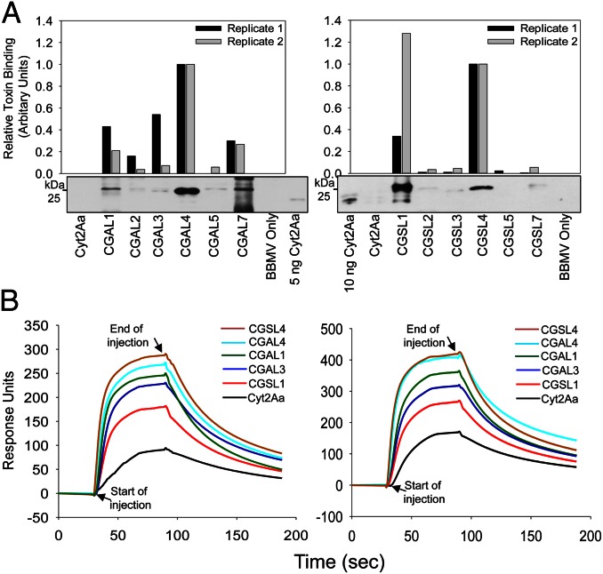
CGALn and CGSLn bind to pea aphid gut BBMV more strongly than Cyt2Aa. (A) Pull-down assays were conducted following incubation of activated Cyt2Aa, CGALn, or CGSLn and pea aphid gut BBMV. Membrane bound toxin was detected by Western blot with anti-Cyt2Aa antiserum. Western blot images (Lower) were scanned and processed using ImageJ to estimate the relative amount of activated toxin associated with pea aphid BBMV (histogram Upper). Relative toxin binding is shown for two pull-down assays with Western blot images shown for Replicate 1 in each case. (B) BIAcore surface plasmon resonance analysis of toxin binding to small unilamellar vesicles (SUV). Sensorgram showing the real-time interaction between 6 µM Cyt2Aa, CGAL1, CGAL3, CGAL4, CGSL1, CGSL4, and immobilized pea aphid gut membrane SUV. L1 chip surfaces were prepared with 4,000 RU of ligands. Data are shown for two independent experiments.

