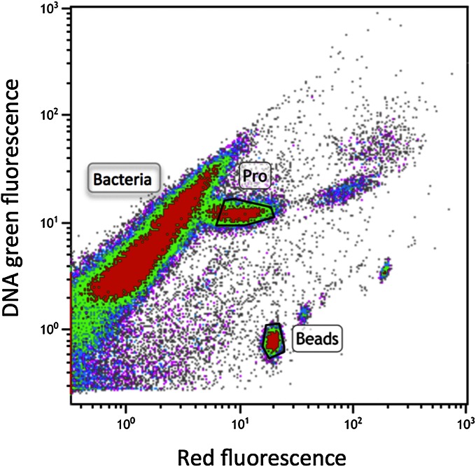
Fig. 5.
Flow cytometric scatter plot showing a signature of SYBR Green I DNA stained picoplankton from station 35 of cruise AMT-21 (47 m depth). Prochlorococcus cells were identified by their extra red chlorophyll fluorescence, gated (area marked “Pro”) and sorted to determine their 3H-glucose uptake.

