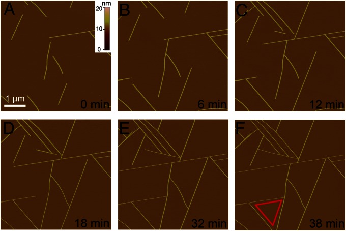
Fig. 2.
In situ AFM observation of the assembly of GAV-9 peptide on mica surface under 100 mM MgCl2. (A–F) Series of snapshots of the extending nanofilaments with a height of 6 nm. The edges of the equilateral triangle in F indicate three preferred directions along which peptides assembled into nanofilaments. The same scale bar used in A applies to all AFM images.

