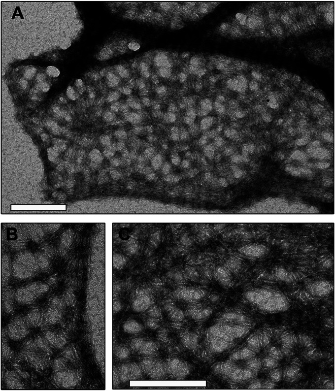
Fig. 1.
FilP forms cis-interconnected networks in vitro. Denatured His-FilP in 8 M urea was slowly dialyzed into 50 mM Tris-buffer pH 7 at +4 °C overnight. Filaments were negatively stained and visualized by transmission electron microscopy. (Scale bars: 200 nm.) (A) View of a larger area covered by networks and compact striated filaments. (B) Shows in higher magnification that a network is a continuous structure with a striated filament. (C) Shows a regular hexagonal pattern and areas with variable compaction in the network.

