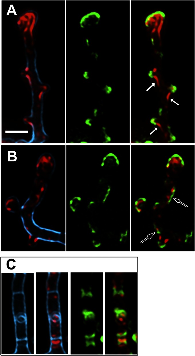
Fig. 5.
DivIVA recruits FilP cytoskeleton. (A–C) Superimposed deconvolved Z-stacks of fluorescence microscopy images of strain K121 after induction of divIVA for 2 h. Blue, cell wall stain (wheat germ agglutinin); green, DivIVA-EGFP fluorescence; red, anti-FilP immunofluorescence. Arrows in A point to filamentous structures of FilP trailing from main hyphae into newly forming branches; arrows in B point to examples of lateral DivIVA signals, not associated with bulges, but associated with enhanced signals of FilP. (C) A hyphal segment with two clearly visible crosswalls (blue), both flanked by DivIVA-GFP and FilP signals. (Scale bar: A, 2 µm; applies to all images.)

