Fig. 2.
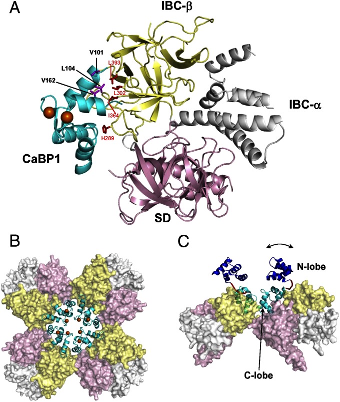
Structure of CaBP1 bound to InsP3R. (A) The C lobe of Ca2+-bound CaBP1 [cyan; Protein Data Bank (PDB) ID code 2K7D] bound to NT (PDB ID code 3UJ4) in a 1:1 complex. Key residues at the binding interface are highlighted in magenta (CaBP1) and red (InsP3R). NT subdomains are colored pink (SD), yellow (IBC-β), and gray (IBC-α). (B) Model of tetrameric NT (pink, yellow, and gray) generated by superimposing the NT crystal structure (29) onto a cryo-EM structure of InsP3R1 (34). NMR structural restraints were used to define contacts between each NT and C lobe of CaBP1 (cyan, with Ca2+ in orange). (C) Side view of tetrameric NT bound to full-length CaBP1 in a 4:4 complex. The N lobe of CaBP1 (blue) is connected to the C lobe (cyan) by a flexible linker (red) that allows the N lobe to adopt multiple orientations (indicated by the arrow).

