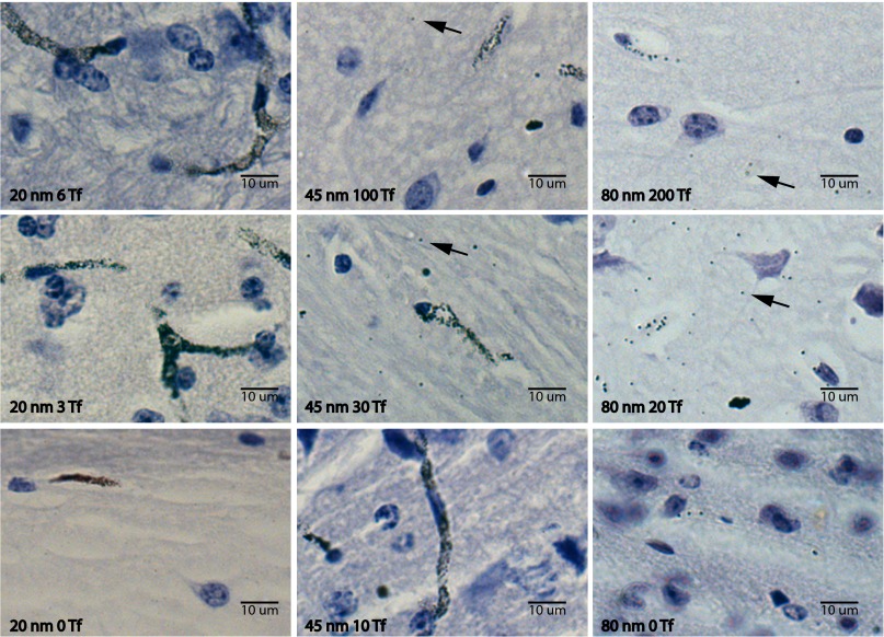
Fig. 4.
Sample images from hematoxylin-stained and silver-enhanced brain sections. Shown are images from a range of nanoparticle formulations injected systemically with brains resected and processed 8 h later. Black arrows accentuate clearly visible nanoparticles. (Left) 20-nm nanoparticles with 6 Tf, 3 Tf, and 0 Tf (from top to bottom). (Center) 45-nm nanoparticles with 100 Tf, 30 Tf, and 10 Tf. (Right) 80-nm nanoparticles with 200 Tf, 20 Tf, and 0 Tf.

