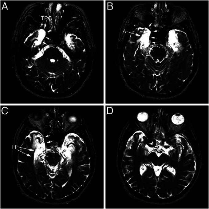
Fig. 1.
T2-weighted MRI images of E.P.’s brain from 1994. Axial sections are arranged from ventral to dorsal (A–D). Note areas of hyperintensity in the medial temporal lobe indicated by white arrows. Regions included within the abnormal hyperintensity include the temporopolar cortex (TPC), amygdaloid complex (A), and hippocampal formation bilaterally (H). The left side of each image illustrates the left side of the brain.

