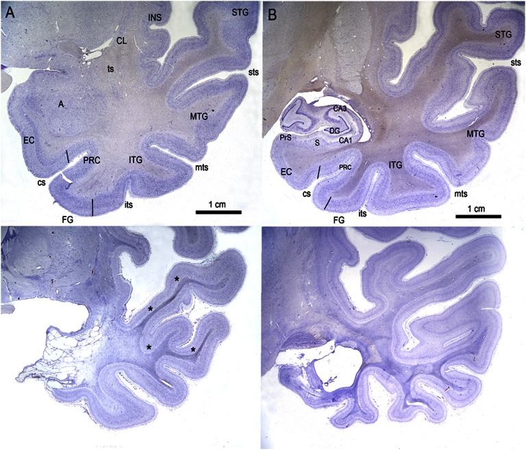
Fig. 7.
(A and B) Higher-magnification photomicrographs of Nissl-stained coronal sections through the medial temporal lobe of E.P. (Lower) and control H.T. (Upper). A is through the level of the amygdaloid complex, and B is through the level of the rostral hippocampal formation. The gliosis and shrinkage of the white matter of the temporal stem and white matter of the superior, middle, inferior, and fusiform gyri are also evident (asterisks). A, amygdala; CA1, CA1 field; CA3, CA3 field; CL, claustrum; cs, collateral sulcus; DG, dentate gyrus; EC, entorhinal cortex; FG, fusiform gyrus; INS, insular cortex; ITG, inferior temporal gyrus; its, inferior temporal sulcus; MTG, middle temporal gyrus; mts, middle temporal sulcus; PRC, perirhinal cortex; PrS, presubiculum; S, subiculum; STG, superior temporal gyrus; sts, superior temporal sulcus; ts, temporal stem.

