
Fig. 1.
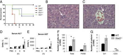
LPS-induced mortality in mice lacking IFN signaling mediators and sepsis-induced tissue inflammation in Stat2−/− mice. (A) Survival curves in WT, Tyk2−/−, Ifnar1−/−, Stat1−/−, and Stat2−/− mice following LPS challenge at a dose of 70 mg/kg. (B and C) Immune cells rapidly accumulate in the extravascular space of livers in Stat2−/− mice compared with WT following LPS challenge. H&E-stained sections of Stat2−/− mouse liver after 9 h LPS challenge (magnification of 600×) show signs of inflammation, microabscesses in liver parenchyma, and leukocytes within hepatic sinusoids (white arrows). (D and E) Serum concentrations (mean ± SEM) of alanine aminotransferase (ALT) and aspartate aminotransferase (AST) from three to five animals following i.p. injection with PBS solution or LPS at time points indicated (*P < 0.05). (F and G) Number of cells in peritoneal lavage fluid of mice after 6 h of LPS (20 mg/kg). Total number (*P = 0.02), F4/80+, GR1+, and CD3+ (**P < 0.001).