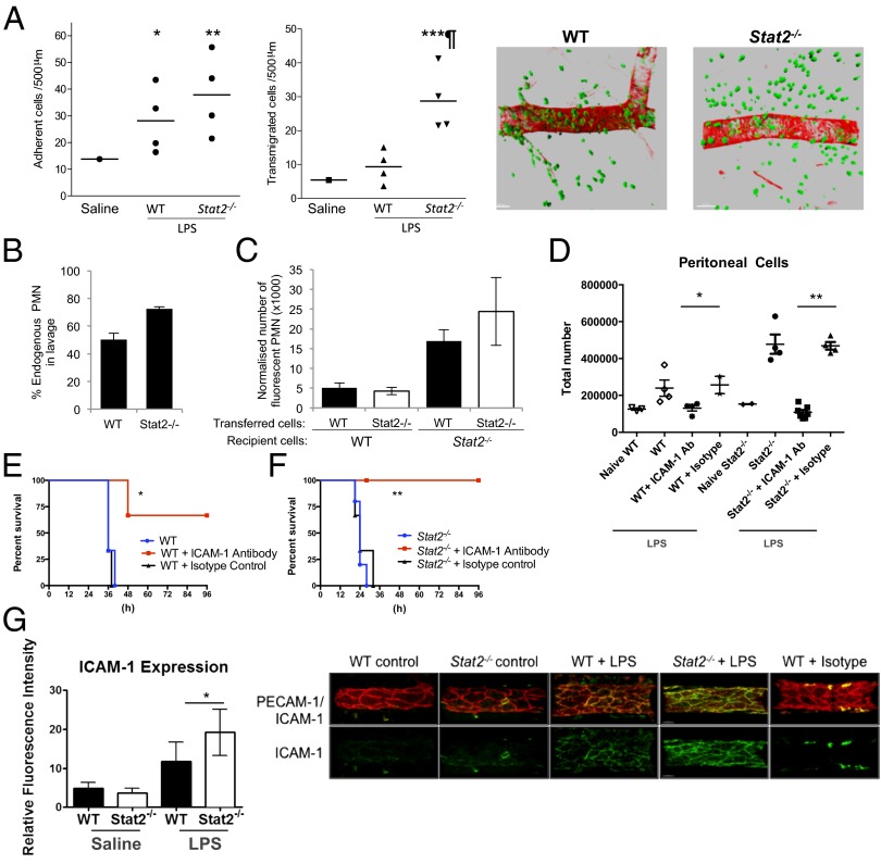
Fig. 3.
LPS-induced death in Stat2−/− mice is mediated by cellular extravasation. (A) WT or Stat2−/− mice received intrascrotal injections of saline solution or LPS (300 ng). Leukocyte responses of adhesion (Left) and transmigration (Right) were quantified 4 h later in cremaster muscle by intravital microscopy (*P < 0.05, **P < 0.01, and ***P < 0.001, saline solution vs. LPS; ¶P < 0.001, WT vs. Stat2−/− mice). Tissue immunofluorescence microscopy for markers of blood vessels (α-smooth muscle actin; red) and neutrophils [myeloid related protein-14 (MRP-14); green]. A representative example of LPS-stimulated WT (Left) and Stat2−/− (Right) tissues. WT and Stat2−/− recipient animals received an adoptive transfer of singly fluorescently labeled primary WT and Stat2−/− bone marrow cells in equal amounts before i.p. administration of LPS (1 mg/kg). Peritoneal lavage was performed 18 h later, and the number and phenotype of WT and Stat2−/− immune cells within the lavage was quantified by FACS analysis. (B) Percentage of endogenous polymorphonuclear neutrophils (PMN). (C) Total number of WT or Stat2−/− fluorescent polymorphonuclear neutrophils retrieved from peritoneal cavities of WT or Stat2−/− mice. (D) WT and Stat2−/− mice received a single dose of ICAM-1 blocking antibody or isotype antibody control 18 h before i.p. LPS challenge (20 mg/kg). ICAM-1 inhibition reduced cellular egress into the peritoneum (*P = 0.026 and **P = 0.0001) and (E and F) survival of mice (*P = 0.02 and **P = 0.007). (G) WT and Stat2−/− mice (n = 6; n ∼ 6 vessels per animal) received LPS intrascrotally (300 ng) for 4 h and ICAM-1 expression was quantified (*P < 0.05). Examples of a maximum intensity projection depicting PECAM1 and ICAM-1 expression (and ICAM-1 isotype control) on WT and Stat2−/− venules are also shown. (Scale bar: 30 μm.)

