Fig. 5.
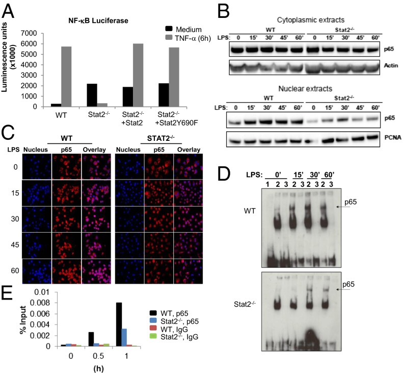
Stat2 deficiency impairs nuclear localization of NF-κB p65. (A) Measurement of NF-κB luciferase reporter activity in WT and Stat2−/− immortalized BMDMs reconstituted with Stat2 or mutant Stat2 (Y689F) and stimulated with TNF-α (10 ng/mL). Data are shown as mean luminescence units (magnification of 1,000×) from two experiments. (B) Immunoblot analysis of NF-κB p65 using nuclear and cytoplasmic extracts from immortalized BMDMs stimulated with LPS (1 μg/mL). (C) LPS induced nuclear translocation of NF-κB p65 (red) assessed by immunofluorescence microscopy. Nucleus is shown in blue. Images are representative of three independent experiments. (D) EMSA of nuclear extracts prepared from LPS-stimulated immortalized BMDMs. Lane 1 is the free probe, lane 2 the labeled probe, and lane 3 the labeled probe competed with 200× molar excess of unlabeled probe. Images shown are representative experiments using at least three independent extracts. (E) Quantitative ChIP assay performed at the indicated time points with antibody against p65 or irrelevant IgG as indicated. Specific quantitative PCR primers were used to amplify the proximal TNF-α promoter region.

