
Fig 1.
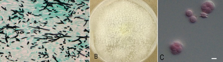
(A) Gomorri methanamine silver-stained section of sinus mucosal tissue showing numerous branched, septate hyphae (bar = 20 μm); (B) colony on OAT showing ascomata after 7 days of growth at 35°C; (C) ascospores in face and side view showing two crests (bar = 5 μm).