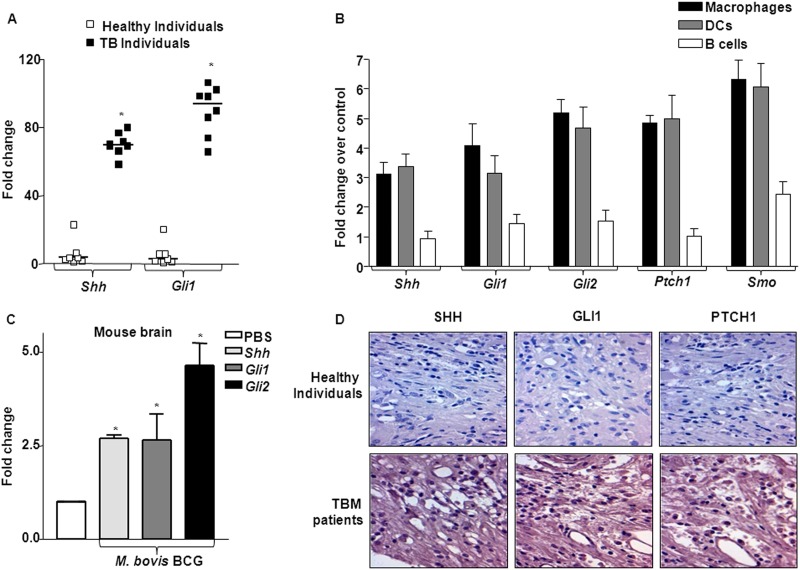
Fig 2.
In vivo induction of SHH signaling. (A) RNA isolated from total PBMCs of healthy individuals (n = 7) or TB patients (n = 7) was used to analyze levels of Shh, Gli1, and Gli2 transcripts. *, P < 0.05 compared to PBMCs of healthy individuals. (B) Macrophages, DCs, and B cells were isolated from total PBMCs of healthy individuals, followed by infection with M. bovis BCG. SHH signaling pathway components were assayed using quantitative real-time RT-PCR (mean ± SE, n = 3). (C) M. bovis BCG bacilli were inoculated into the cranial cavity of one set of mice (n = 4), while the other set received PBS as a mock infection. After 5 days of infection, mice were sacrificed and total RNA from brain tissues was isolated to investigate the status of SHH signaling using quantitative real-time RT-PCR. *, P < 0.05 versus mice receiving PBS. (D) Brain tissue sections obtained from healthy individuals or TB meningitis patients were stained with polyclonal antibodies against SHH, GLI1, and PTCH1 proteins. Sections were counterstained with hematoxylin-eosin.

