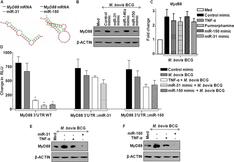
Fig 8.
SHH signaling-responsive miR-31 and miR-150 target MyD88 to suppress TLR2 signaling. (A) Hybridization analysis of 3′ UTR of MyD88 with mature oligonucleotide sequences of miR-31 and miR-150 using RNAhybrid software. The structures with minimum free energy are shown. (B) miR-31, miR-146a, and miR-150 were overexpressed in macrophages by transfecting respective mimics, and levels of MyD88 expression were assayed by immunoblotting using anti-MyD88 antibody. Data are representative of 3 independent experiments. (C) The level of Myd88 transcript expression was monitored under the indicated conditions by real-time quantitative RT-PCR using Myd88-specific primers. Data represent means ± SEs (n = 3). (D) THP1-derived macrophage cells were transfected with the WT MyD88 3′ UTR luciferase construct or MyD88 3′ UTR ΔmiR-31 or MyD88 3′ UTR ΔmiR-150 luciferase constructs along with miR-31 or miR-150 mimics, as indicated. At 72 h posttransfection, TNF-α treatment or M. bovis BCG infection was done as shown and luciferase assay was performed. Data represent means ± SEs (n = 3). RLU, relative light units. *, P < 0.05 versus control. (E and F) The level of MyD88 protein in macrophages was assessed either with TNF-α treatment or with overexpression of miR-31 (E) or miR150 (F). Results of a representative MyD88 immunoblotting analysis of 3 separate experiments are shown.

