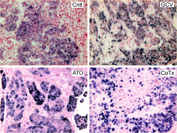
Figure 2.
CNE1 < BX1 > cells within the tumors display EBER positivity and are morphologically distinct from mouse cell infiltrates. In sections of tumor explants 22 days post injection, EBER-1 and EBER-2 were detected by cytogenic in situ hybridization and stained with NBT/BCIP (blue). Nuclei were stained with Nuclear Fast Red counterstain (original magnification of 200×).

