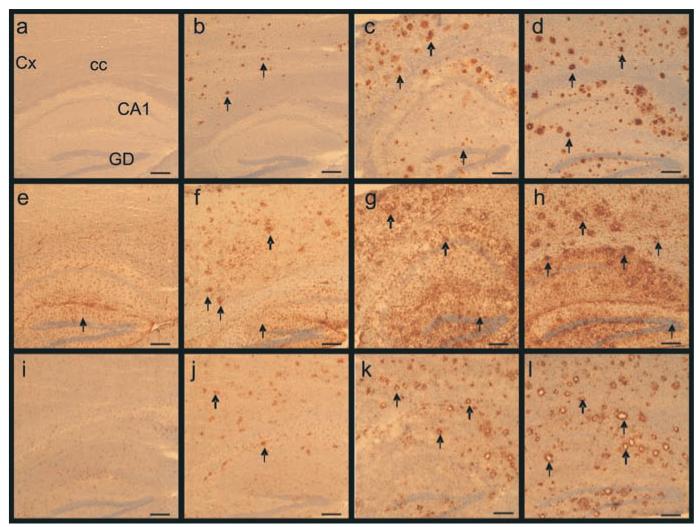
Figure 6.
Increased number and size of amyloid deposits, associated astrocytosis and microgliosis with aging in APP/PS1 mice. (a-d) Staining for amyloid deposits (arrows) in the cortex and the hippocampus of 3, 6 and 12-month old APP/PS1 mice (b, c, d respectively). (a) shows the lack of staining in a 12-month-old control (C57/Bl6) mouse (4G8 antibody). (e-h) Reactive astrocytes (arrows) in the brain of a 12-month-old control (C57/Bl6) mouse (e) and 3 (f), 6 (g), 12-month-old (h) APP/PS1 mice (GFAP antibody). Microgliosis (arrows) in the brain of 3 (j), 6 (k), and 12-month-old (l) APP/PS1 mice (IBA1 antibody). No staining could be detected in control mice (i). Annotations: Cx: Cortex; cc: Corpus callosum; CA1: CA1 region of the hippocampus; DG: Dentate gyrus of the hippocampus. Scale bars= 200μm.

