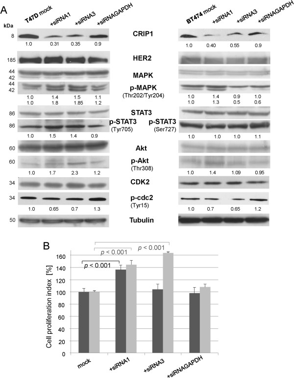
Figure 4.
CRIP1 silencing results in the activation of signaling proteins involved in cell proliferation. (A) Western blot analysis showing the expression and phosphorylation levels of signaling proteins after the knockdown of CRIP1 in T47D and BT474 breast cancer cells using HER2, (phospho) MAPK, (phospho) STAT3, (phospho) Akt and (phospho) cdc2 antibodies. Tubulin was used as a loading control. The mean values of three independent experiments are shown. (B) Seventy-two hours after transfection, the WST-1 reagent was added to a defined amount of T47D and BT474 cells and the absorbance measured after 3 h is shown. The graph represents the amount of viable cells in relation to the mock control. The means of five independent experiments, the standard deviations and the p-values are shown. For statistical analyses, the student´s t-test was performed.

