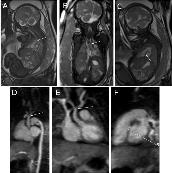
Figure 3.
A 34-week fetus, interruption of the aortic arch with VSD. Fetal MR FIESTA oblique sagittal and coronal view images show the interruption of the aortic arch (arrows in A and B). MR FIESTA short-axis view image shows the VSD (arrow in C). Postnatal follow-up contrast-enhanced magnetic resonance angiography (CE-MRA) oblique sagittal and coronal view images show the interruption of the aortic arch (arrows in D and E). Postnatal CE-MRA short axis view image shows the VSD (measuring mark in F), compared to prenatal imaging.

