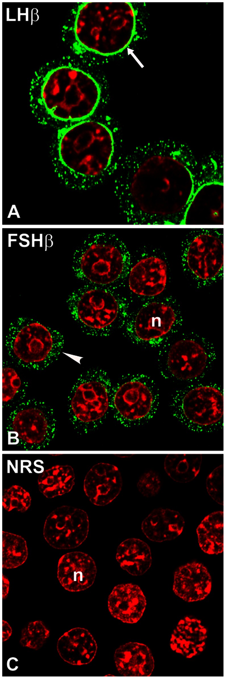
Figure 2. Subcellular localization of LHβ (A) and FSHβ (B) subunit in GH3 cells.

The cells were immunostained with CGβ antiserum (green) and monoclonal antibody against FSHβ subunit (green). Note unique ER/perinuclear staining pattern for LHβ (A, arrow) vs. dispersed cytoplasmic puncta for FSHβ subunit (B, arrowhead). The n indicates the nucleus (red). The micrographs shown are representative of four to eight experiments and are at the X100 and X150 magnification. NRS (C), normal rabbit serum.
