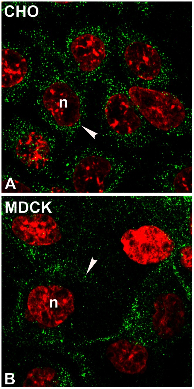
Figure 6. Immunostaining of LHβ subunit in CHO (A) and MDCK (B) cells.

The cells were immunoprobed with CGβ antiserum (green). Note that LHβ shows dispersed cytoplasmic puncta (A, B, arrowhead) with no ring-like pattern near nucleus. The n indicates the nucleus (red). The micrographs shown are representative of four experiments. X150.
