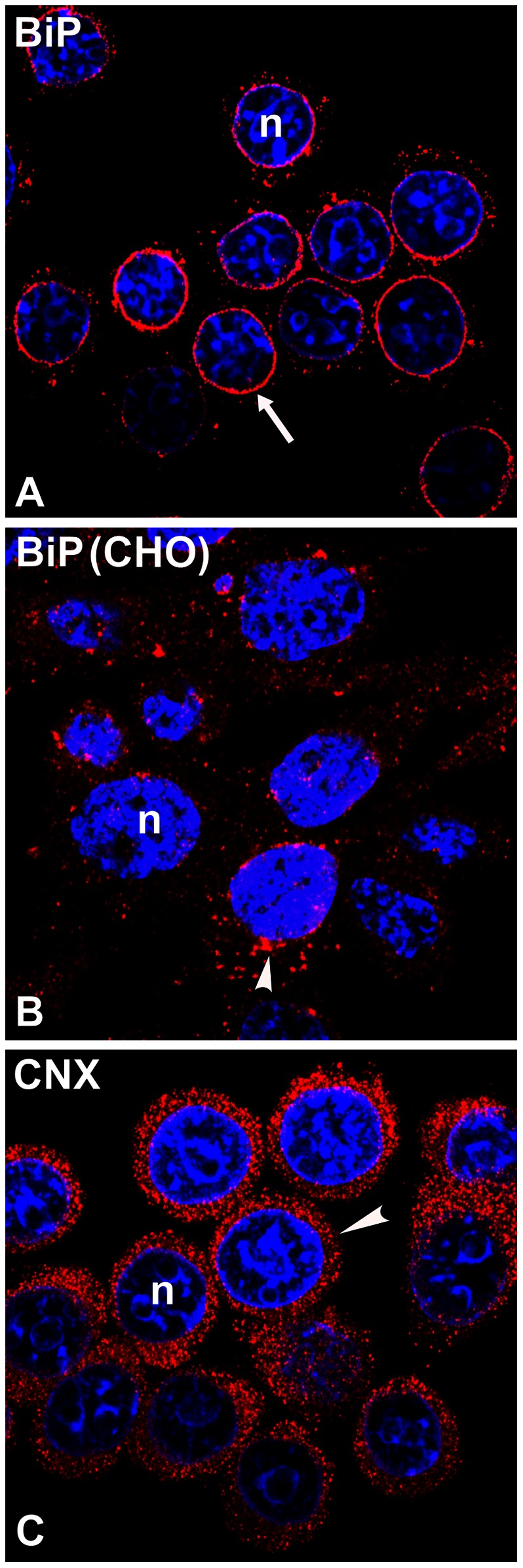
Figure 7. Immunolocalization of endogenous BiP (A, B) and calnexin (CNX, C) in non-transfected GH3 or CHO cells.

For GH3 cells the BiP antiserum (A, red) stained predominantly around nuclei (arrow), while the CNX antiserum (C, red) showed peripheral ER staining (arrowhead). Note that BiP in CHO cells (B) is localized as dispersed cytoplasmic puncta with some aggregation near the NE (arrowhead). Nuclei (n) were counterstained using TOPRO-iodide-3 (blue). The micrographs shown are representative of four experiments.
