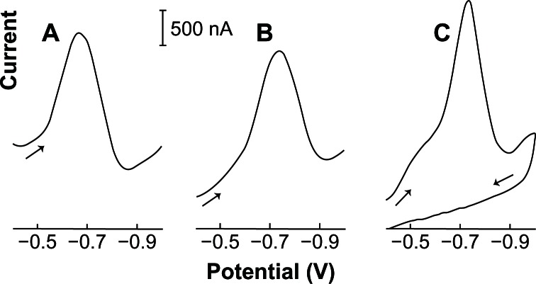
Figure 2.
Differential-pulse (A), linear-scan (B), and adsorptive linear cyclic (C) voltammograms of 0.05 ppm acyclovir in 2.0 × 10⁻³ mol L−1 NaOH (with 1% v/v of ethylic alcohol). Condition time, 60 seconds at −0.90 V; accumulation time, 120 seconds at −0.40 V with stirring; amplitude pulse, 50 mV (A); scan rate, 50 (A), 100 (B and C) mV s−1; thin-film mercury electrode (5 minutes at −0.9 V).

