Figure 2.

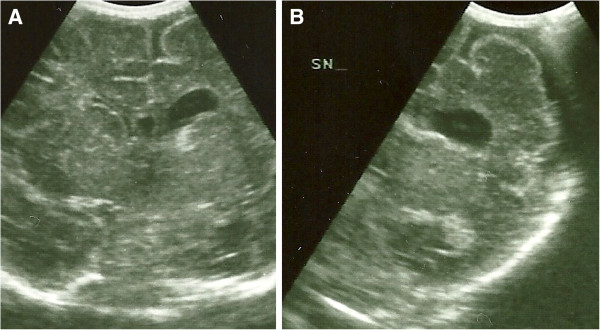
Neonatal cerebral ultrasonographic coronal images. A Left ventriculomegaly, B Left hemisphere atrophy with enlarged subarachnoid spaces.

Neonatal cerebral ultrasonographic coronal images. A Left ventriculomegaly, B Left hemisphere atrophy with enlarged subarachnoid spaces.