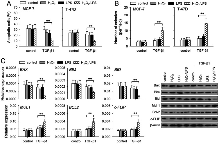
Figure 5. TGF-β1/H2O2/LPS promotes anoikis-resistance of non-invasive breast cancer cells.
MCF-7 and T-47D cells were cultured for 8 days in absence or presence of TGF-β1 (5 ng/ml), H2O2 (50 µM), and LPS (100 ng/ml). The cells were then used in following experiments. (A) The cells were transferred to poly-HEMA-coated plate and cultured for 24 h. The apoptosis of the cells was analyzed by flow cytometry. (B) The cells were then cultured in soft agar for 3 weeks. The colonies were counted. (C) MCF-7 cells were used for the assay of the expression of Bax, Bim, Bid, Bcl-2, Mcl-1 and c-FLIP. The expression of these genes was detected by real-time RT-PCR and Western blot. P values, *P<0.05, **P<0.01.

