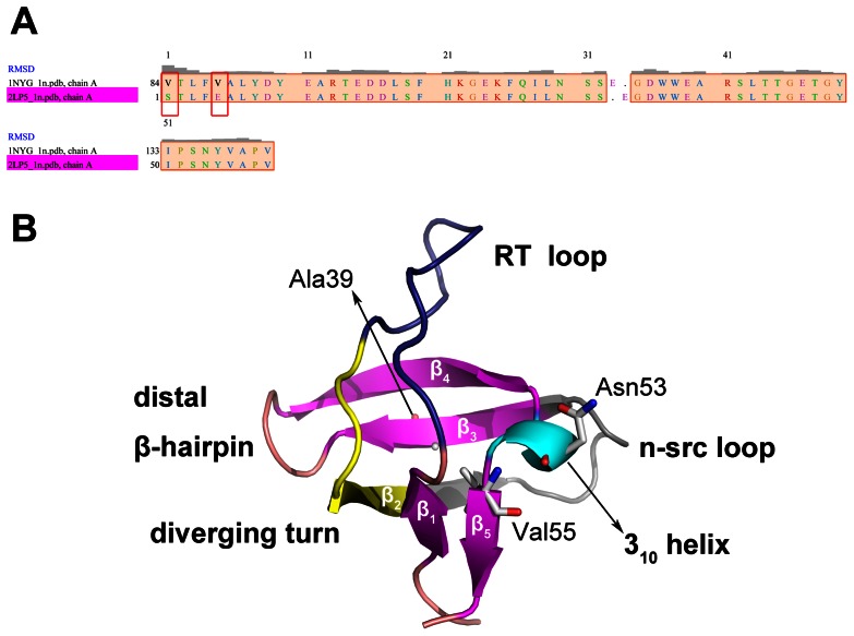
Figure 1. SH3 domain sequence and structure.
Sequence alignment of the human (1NYG) and gallus (2LP5) SH3 domain (A) is generated by Chimera 1.5.3 [42]. Only residue 1 and residue 5 between the two sequences are different. In crystal structure of the gallus SH3 domain (PDB ID: 2LP5) (B), the SH3 domain is divided into several parts, such as RT loop, diverging turn, N-src loop, distal β-hairpin, 310 helix, and five strands. Mutation sites, Ala39, Asn53, and Val55 are shown in white sticks and labeled in the figure. All the oxygen atoms and nitrogen atoms of these residues are colored red and blue, respectively.

