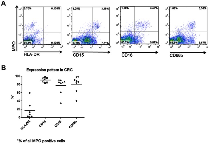
Figure 2. Phenotypic characterization of CRC infiltrating MPO+ cells.
CRC surgical specimens were enzymatically digested and immediately stained with fluorochrome labeled mAbs recognizing MPO, HLA-DR, CD66b, CD15 and CD16, as indicated in “materials and methods”. Panel A reports one representative staining, whereas panel B summarizes results from freshly excised specimens (n = 8) regarding the expression of the indicated markers in CRC infiltrating MPO+ cells.

