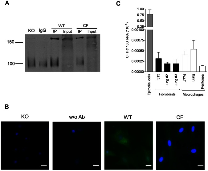
Figure 3. Mouse fibroblasts express CFTR protein.
CFTR expression in mouse nasal epithelial cells in primary cultures, fibroblasts (3T3 cell line and lung cells at passages #2 and #3) and macrophages (J774 cell line and alveolar and peritoneal cells in primary cultures). a) Immunoblots of precipitates for IgG blank and lung fibroblasts from Cftr knockout mice (KO), wild-type (WT) and CF mice homozygous for the F508del mutation. Immunoprecipitates (IP) using 4×10⁶ fibroblasts grown in Petri dishes were lysed in a buffer (25 mM Tris-HCl pH 7.5, 150 mM NaCl, 1% Triton X-100) supplemented with Complete PIC (Roche) and incubated with mouse anti-CFTR antibody clone 24-1 coupled with G protein-conjugated magnetic Dynabeads. Data selected from at least 4 experiments with similar results. As expected for mouse CFTR, a band was recognized at 160 kDa but not detected when IPs were performed with non-immune IgG. b) Immunofluorescence labelling for CFTR (green) in fibroblasts purified from Cftr knockout (KO) mice, wild-type mice with (WT) or without CFTR antibody (w/o Ab), and CF mice homozygous for the F508del mutation. Fibroblasts grown on collagen-coated cover glasses were fixed with acetone and permeabilized with 0.25% Triton X-100. Detection was obtained using anti-mouse AlexaFluor 488 secondary antibody after overnight incubation with anti-CFTR antibody clone 24-1. Nuclei stained with DAPI blue. Bars: 20 µm. Data selected from at least 3 experiments with similar results. c) Total CFTR mRNA expression, using 18S rRNA as a reference. Data expressed as means ± SEM of 3–8 samples per group.

