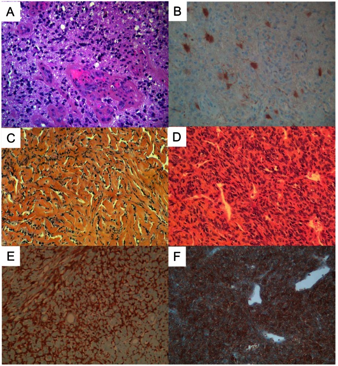
Figure 4. Microscopic aspects and ALDH1 expression using IHC.
A. Microscopic features (HES) of a glioblastoma used as positive control for ALDH1 immunostaining. B. ALDH1 expression in the cytoplasm of few astrocytic tumor cells. C–D. Microscopic features (HES) of an SFT in a collagenic area (C) and of an SFT in a cellular area with an “hemangiopericytoma” vascular pattern (D). E–F. ALDH1 immunostaining in a collagenic area (E) and in a cellular area (F): note the strong and diffuse expression in the cytoplasm of tumor cells. For all images, magnification is ×25.

